- Share
- Share on Facebook
- Share on X
- Share on LinkedIn
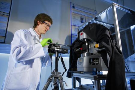
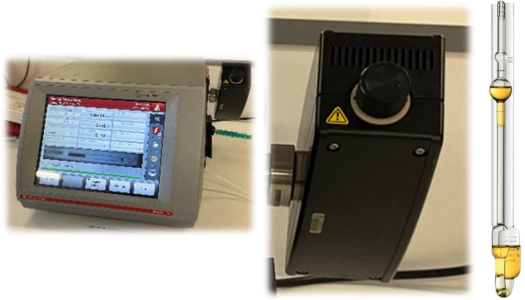
The Rheometry and Coupled Physical Measurement Platform of the Rheology and Processes Laboratory brings together various instruments that allow for detailed observation of materials in flow. We have rotational rheometers, a high-frequency rheometer, a viscometer, an extensional rheometer, techniques of microrheometry and also light diffusion benche to study the rheological behavior of materials and their structure.
Rotational Rheometry
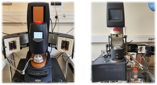
We have a wide range of rotational rheometers operating under controlled stress or strain, along with a variety of accessories.
ARES-G2
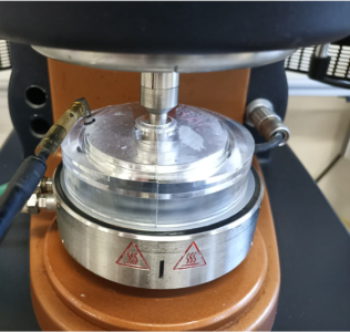
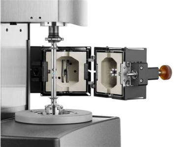
Our strain imposed rheometer ARES-G2 is equipped with a high-temperature oven and a connection to liquid nitrogen, allowing it to perform rheological measurements between -150 and 600°C, or under neutral gas. It is also equipped with a PPS geometry kit for aggressive samples. The DMA mode of the ARES-G2 also enables the testing of a sample with oscillations of up to 50µm amplitud at frequencies of up to 100Hz.
MCR501
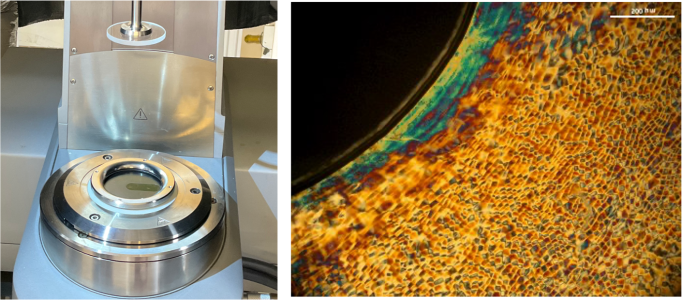
This rheometer is equipped with the Anton Paar rheo-microscope accessory, allowing for observation of the sample under shear with magnifications ranging from x5 to x50. We also have modules for fluorescence studies and polarized light examination.
MARS III
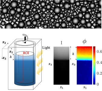
Versatile in nature, it is equipped with a rough large-gap Couette geometry, allowing for the study of charged fluids. A custom-made transparent Couette geometry is also available, enabling observation of sedimentation during measurements.
MCR301
This highly sensitive rheometer is equipped with a 'Mooney Ewart' type geometry, mounted on an optical table and furnished with the necessary accessories for observing 'speckles'.
DHR3
Ce rhéomètre polyvalent permet d’obtenir rapidement une courbe d’écoulement ou toute autre mesure standard. Il est équipé d’un module Couette thermostaté et de géométries rugueuses pour les échantillons glissants.
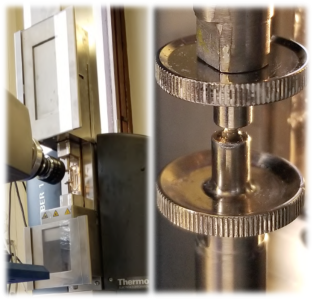
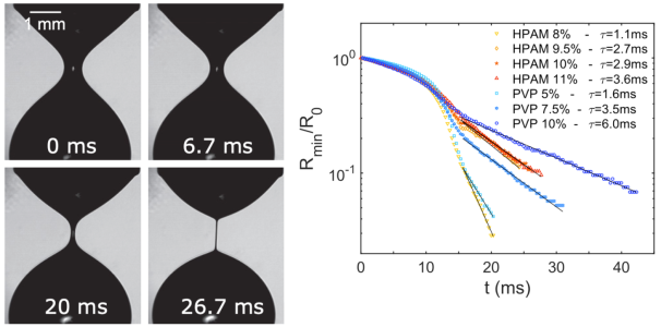
Extensional Rheometry
Light Diffusion
Static ligth scattering (SALS)
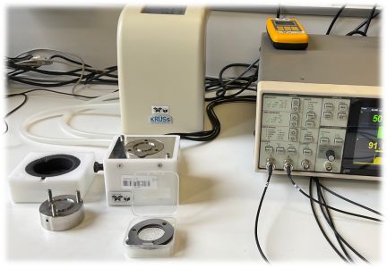
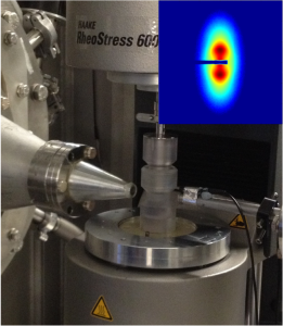
The small-angle light scattering device was developed at the LRP and can be coupled with shear flow, filtration and elongation cells to simultaneously apply external fields to the structural characterisation of the sample.
It consists of a 2 mW laser beam (He-Ne) with a wavelength of 632.8 nm and a Fresnel lens to collect the scattered light (Piau et al., 1999). The detector is an Allied Vision monochrome digital camera (AV MAKO G-419B POE): CMOS sensor (2048 x 2048 pixels, 11.3 x 11.3 mm2). The images were processed using a video system and specially developed software (Vimba Matlab) that enables the scattered intensity to be grouped and averaged according to different groupings (radial, angular sectors, corona) using SAXS Utilities software (Sztucki and Narayanan, 2007). The scattering spectra are recorded by the camera and video system throughout the experiment. The light scattering measurements cover a range of wave vectors (q) between 2 × 10⁻⁴ and 4 × 10⁻³ nm-1.
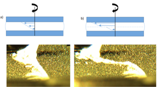
In the example illustrated below, a shear flow cell consists of a rectangular quartz channel, as shown in the diagram below. The shear flow cell is coupled to a syringe pump to vary the flow rate Q and therefore the shear rate inside the channel, typically from 10-2 to 1000 s-1.
References
Piau, J.M., Dorget, M., Palierne, J.F., Shear elasticity and yield stress of silica–silicone physical gels: Fractal approach, J. Rheol., 43, 305–314 (1999).
Pignon, F., Piau, J.M. and Magnin, A., Structure and pertinent length scale of a discotic clay gel, Physical Review Letters, 76, 4857-4860 (1996). https://link.aps.org/doi/10.1103/PhysRevLett.76.4857
Pignon, F., Magnin, A. and Piau, J.M., Butterfly light scattering pattern and rheology of a sheared thixotropic clay gel, Physical Review Letters, 79, 4689-4692 (1997). https://link.aps.org/doi/10.1103/PhysRevLett.79.4689
Pignon, F., Magnin, A., Piau, J.M., Cabane, B., Lindner, P. and Diat, O., A yield stress thixotropic clay suspension: investigations of structure by light, neutron and x-ray scattering, Physical Review E, 56, 3281-3289 (1997). https://link.aps.org/doi/10.1103/PhysRevE.56.3281
Pignon, F., Magnin, A. and Piau, J.M., Thixotropic behavior of clay dispersions: combinations of scattering and rheometric techniques, Journal of Rheology, 42, 1349-1373 (1998). https://doi.org/10.1122/1.2079267
Saint-Michel F., Pignon F. and Magnin A., Fractal behavior and scaling law of hydrophobic silica in polyol, Journal of Colloid and Interface Science, 267(2) 314-319 (2003).
de Bruyn J.R., Pignon F., Tsabet E. and Magnin A. Micron-scale origin of the shear-induced structure in Laponite–poly(ethylene oxide) dispersions, Rheologica Acta, 47, 63-73 (2008).
Fernández V.A., Tepale N., Alvarez J.G., Pérez-López J.H., Macías E.R., Bautista F., Pignon F., Rharbi Y., Gámez-Corrales R., Manero O., Puig J.E. and Soltero J.F. A., Rheology of the pluronic P103/water system in the semidilute regime: evidence of non-equilibrium critical behavior, Journal of Colloid and Interface Science, 336, 842-849 (2009).
Pignon, F., Challamel, M., De Geyer, A., Elchamaa, M., Semeraro, E.F., Hengl, N., Jean, B., Putaux, J.L., Gicquel, E., Bras, J., Prevost, S., Sztucki, M., Narayanan, T., Djeridi, H., Breakdown and buildup mechanisms of cellulose nanocrystal suspensions under shear and upon relaxation probed by SAXS and SALS, Carbohydrate Polymers, 260, 117751 (2021).
Bauland J., Andrieux V., Pignon F., Frath D., Bucher C., Gibaud T., Viologen-based supramolecular crystal gels: gelation kinetics and sensitivity to temperature, Soft Matter, 20, 8278, (2024).
Mandin S., Metilli L., Karrouch M.,Blésès D., Lancelon-Pin C., Sailler P., Chèvremont W., Paineau E., Putaux J.L., Hengl N., Jean B., and Pignon F., Multiscale study of the chiral self-assembly of cellulose nanocrystals during the frontal ultrafiltration process, Nanoscale, 16, 19100, (2024). https://doi.org/10.1039/D4NR02840F
Support references
Sztucki M., Narayanan T., Development of an ultra-small-angle X-ray scattering instrument for probing the microstructure and the dynamics of soft matter, J. Appl. Cryst., 40, 459–462 (2007).
Son, Y. Determination of shear viscosity and shear rate from pressure drop, and flow rate relationship in a rectangular channel, Polymer, 48, 632–637 (2007).
Microrheology
Passive microrheology
Passive microrheology consists in quantifying the Brownian motion of micro-particles in a medium whose rheological properties are to be characterized.
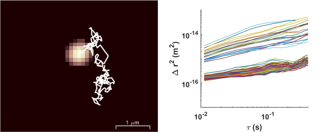
Confocal image of a liquid containing fluorescent tracer particles (diameter 0.5 µm) and magnified view of a single tracer particle and its Brownian trajectory.
The analysis of particle motion makes it possible to calculate their mean square displacement (MSD). In Newtonian liquids, viscosity can be measured over a wide range (from 10⁻³ Pa·s to 10 Pa·s). In purely elastic systems, the elastic modulus can be measured within a range of 0.1 to 20 Pa. The accessible frequency range extends from 0.1 to 20 Hz.
- The advantages of passive microrheology include:
- The very small sample volumes required,
- the ability to analyze heterogeneous samples,
- in-situ monitoring of rheological properties.
Example of microrheology results on gel samples [D. Milian et al., Biomacromolecules 2023]. As prepared the gel is purely elastic and the MSDs are uniform and time-independent. After a pre-shear, microcracks are visible and tracer particles located in these microcracks exhibit much higher MSD and a diffusive behavior.
- Share
- Share on Facebook
- Share on X
- Share on LinkedIn